

On this page
Executive Summary
Periodontal disease is the most common preventable health condition affecting companion animals, affecting over 80% of dogs and 70% of cats older than 3 years of age. Despite its prevalence, dental disease remains significantly undertreated in veterinary practice, particularly in resource-limited settings like India where preventive care adoption is low (18% of pet owners).
This guide provides comprehensive, evidence-based information on:
- Epidemiological trends in companion animal dentistry
- Detailed pathophysiology of disease progression
- Clinical classification and diagnostic protocols
- Systemic complications and health impacts
- Professional treatment standards and outcomes
- Evidence-based prevention strategies
Key clinical insight: Early intervention in stage 1 (gingivitis) is 90% reversible, while advanced stages (periodontitis) result in permanent tissue damage requiring ongoing management. Prevention and early detection are cost-effective and significantly improve long-term outcomes.
Epidemiology & Prevalence
Global Disease Burden
Canine Periodontal Disease
Prevalence Data:
- 80% of dogs over age 3 years show signs of periodontal disease
- 90% of dogs over age 4 years have documented dental pathology
- Prevalence increases progressively with age:
Ages 1-3 years: 20-30% prevalence
- Ages 3-7 years: 60-80% prevalence
- Ages 7+ years: 85-95% prevalence
Breed Predisposition:
- Toy breeds (<6 kg): Highest prevalence (60-80%)
Chihuahuas: 75% prevalence
- Shih Tzus: 72% prevalence
- Poodles: 68% prevalence
- Small breeds (6-15 kg): 55-65% prevalence
- Medium/Large breeds (>15 kg): 40-50% prevalence
Biological Basis for Breed Predisposition:
- Crowded dentition in toy breeds increases biofilm retention
- Anatomically narrower interdental spaces
- Higher bacterial load concentration
- Reduced masticatory force (less mechanical cleaning)
- Greater genetic predisposition to immune response abnormalities
Feline Periodontal Disease
Prevalence Data:
- 70% of cats over age 3 years show oral pathology
- 90% of cats over age 10 years have dental abnormalities
- Unique feline condition: Odontoclastic Resorptive Lesions (FORL)
Affects 40-60% of domestic cats
- Etiology not fully understood (possibly immune-mediated)
- Often accompanies periodontal disease
Feline-Specific Characteristics:
- Cats often under-diagnosed due to intraoral lesion location
- Owner lack of awareness (cats less likely to show obvious signs)
- Predilection sites: Molars and premolars (particularly line angle)
- Pain behavior often attributed to aging rather than dental disease
India-Specific Epidemiology
Urban Pet Population Data:
- 35% of urban pet dogs in major Indian cities (Mumbai, Delhi, Bangalore, Chennai) show dental abnormalities
- SKS Veterinary Hospital clinical audit: 78% of dogs presenting for general checkup show some degree of periodontal disease
- 40% of pet cats in India carry excess weight and dental disease burden
Preventive Care Gap:
- Only 18% of Indian pet owners provide annual professional dental cleaning
- 90%+ of households feed pets human food regularly (primary risk factor)
- Limited access to veterinary dental specialists outside major metropolitan areas
- Cost barriers (preventive care perceived as luxury rather than necessity)
Unique Risk Factors in Indian Context:
- Dietary Practices:
Biryani, curry, paratha feeding (high fat, starch content promotes bacterial growth)
- Rice and wheat-based scraps (plaque-promoting)
- Spiced foods (gum irritation)
- Inadequate access to commercial dental-specific kibble
- Awareness Barriers:
Misconception that dental disease is “normal aging”
- Limited understanding of systemic complications
- Historical underemphasis on preventive dentistry in veterinary training
- Economic Factors:
Professional cleaning costs (₹5,000-₹12,000) perceived as expensive
- Emergency extraction costs (₹20,000-₹50,000+) still higher
- Limited insurance coverage for preventive care
Pathophysiology & Disease Progression
Bacterial Biofilm Formation & Development
Timeline & Microbial Succession
Phase 1: Initial Colonization (Hours 0-24)
Mechanism:
- Pellicle formation (selective protein deposition on enamel surface)
- Salivary glycoproteins and antibodies form protective matrix
- Initial bacterial colonization (primary colonizers):
Streptococcus sanguinis
- Streptococcus gordonii
- Actinomyces naeslundii
- Actinomyces viscosus
Clinical Features:
- Biofilm thickness: <10 micrometers
- Plaque color: Clear to faint white deposit
- Location: Primarily at gumline
- Visibility: Requires disclosing agent for detection
- Bacterial density: ~10^7 cells/mm³
Reversibility: 100% – can be removed with mechanical cleaning
Phase 2: Early Biofilm Development (Days 1-7)
Microbial Changes:
- Ecological succession begins
- Streptococcus species dominate (gram-positive cocci)
- Actinomyces species increase
- Early anaerobic microniches forming
- Biofilm matrix production increases
Pathophysiology:
- Oxygen tension decreases (anaerobic conditions developing)
- Polysaccharide matrix production (exopolysaccharide accumulation)
- Cell-to-cell communication (quorum sensing) initiation
- Antibiotic tolerance developing (biofilm protection)
Clinical Features:
- Plaque thickness: 50-100 micrometers
- Appearance: White/yellow deposit at gumline
- Adherence: Increasingly difficult to remove
- Bacterial density: ~10^8 cells/mm³
- Gingival response: Minimal or early inflammation
Reversibility: 95% with mechanical intervention
Phase 3: Pathogenic Shift (Days 7-21)
Critical Transition: This phase marks the shift from commensal to pathogenic community.
Gram-Negative Anaerobic Bacteria Emergence:
- Porphyromonas gingivalis (key pathogenic species)
- Prevotella intermedia
- Tannerella forsythia
- Treponema denticola
- Fusobacterium nucleatum
Virulence Factor Production Begins:
- Protease synthesis (tissue destruction)
- Lipopolysaccharide (LPS) production
- Collagenase and hyaluronidase release
- Fibrinolytic enzyme production
- Immunosuppressive molecule generation
Mineralization & Tartar Formation:
- Plaque begins hardening into tartar (calculus)
- Calcium and phosphate deposition
- Mineralization process: 2-7 days after calculus nucleation
- Tartar composition: 70-80% inorganic, 20-30% organic
Clinical Features:
- Visible yellow-brown crusty deposits
- Tartar location: Initially supragingival, extends subgingivally
- Gingival inflammation: Obvious redness at gumline
- Bleeding: Present on probing (gingival ulceration)
- Halitosis: Detectable (volatile sulfur compounds produced by anaerobes)
Reversibility: 85% with aggressive intervention (professional cleaning + antibiotics)
Phase 4: Established Periodontitis (Weeks 3-8)
Disease-Promoting Biofilm Characteristics:
- Complex multispecies consortia (20-30+ species)
- Highly organized architecture (tower-like structures)
- Protected microenvironments (oxygen gradients)
- Active virulence factor production
- Chronic inflammatory infiltrate adjacent
Subgingival Biofilm Development:
- Extension below gumline begins
- Anaerobic environment established
- Gram-negative bacteria proliferation
- Reduced oxygen tension (100 micrometers oxygen penetration)
- Unique subgingival flora (different from supragingival)
Host Inflammatory Response:
- T-lymphocyte infiltration increases
- B-lymphocyte activation
- Cytokine production: TNF-α, IL-1β, IL-6, IL-8
- Polymorphonuclear leukocyte (PMN) recruitment
- Gingival ulceration occurs
Gingival Changes:
- Redness: Capillary dilation
- Swelling/edema: Inflammatory cell infiltration
- Bleeding: Ulcerated epithelium
- Increased gingival crevicular fluid (GCF)
- GCF contains: Inflammatory mediators, bacterial antigens, tissue degradation products
Clinical Features:
- Tartar visible above and below gumline
- Gingival pocket formation (4-6mm probing depth)
- Visible gingival recession starting
- Spontaneous bleeding or bleeding with minimal probing
- Moderate to severe halitosis
Reversibility: 60% with professional cleaning + home care + medical management
Phase 5: Advanced Periodontitis (Months 1-3+)
Progressive Tissue Destruction:
- Apical migration of epithelial attachment
- Periodontal ligament degradation
- Alveolar bone loss initiates (2-3mm annually if untreated)
- Cementum exposure
- Root surface colonization by pathogenic biofilm
Bone Loss Mechanism:
- Osteoclast activation (triggered by inflammatory mediators)
- IL-1β and TNF-α stimulate bone resorption
- Alveolar bone height reduction
- Radiographic changes visible (bone loss appears as radiolucency)
Bacterial Translocation:
- Ulcerated gingival epithelium allows bacterial entry
- Transient bacteremia: Brief bacterial presence in bloodstream
- During chewing: Bacteria-laden fluid enters blood vessels
- During professional cleaning: Temporary bacteremia documented
- Healthy immune system clears bacteria within minutes
Tooth Mobility Development:
- Periodontal ligament destruction reduces attachment
- Grade 1: Slight mobility (<1mm horizontal movement)
- Grade 2: Moderate mobility (1-2mm movement)
- Grade 3: Severe mobility (>2mm, may be mobile mesiodistally AND gingivolabially)
Clinical Features:
- Severe halitosis
- Visible gum recession
- Periodontal pockets: 6-8mm probing depth
- Tooth mobility: Grade 1-2
- Possible suppuration (pus drainage)
- Visible tartar, both supra- and subgingival
- Bone loss: 25-50% on radiographs
Reversibility: 30% – damage partially irreversible; management is long-term
Phase 6: Severe/End-Stage Periodontitis (Months 3+ untreated)
Irreversible Tissue Changes:
- Extensive alveolar bone loss (>50% of attachment)
- Severe periodontal ligament destruction
- Root surface resorption begins
- Tooth mobility: Grade 3 (near exfoliation)
- Potential for spontaneous tooth loss
Systemic Bacterial Translocation:
- Chronic bacteremia may develop (low-level, continuous)
- Repeated bacterial seeding to distant organs
- Immune complex deposition possible
- Chronic inflammatory state systemic
Pulpal Involvement Possible:
- Retrograde pulpitis (infection ascending through apex)
- Pulpal necrosis
- Tooth becomes non-vital (dead tooth)
- Root canal infection
Clinical Features:
- Extreme halitosis
- Severe gum inflammation, possible suppuration
- Probing depth: >8mm
- Tooth mobility: Grade 3
- Exposed roots (gingival recession advanced)
- Possible facial swelling (abscess formation)
- Systemic signs may emerge: Fever, malaise, anorexia
Reversibility: <10% – primarily palliative/extractive management
Key Pathogenic Bacterial Species
Porphyromonas Gingivalis (Primary Pathogen)
Characteristics:
- Gram-negative anaerobic rod
- Black-pigmented colonies (contains porphyrin compounds)
- Found in 90% of canine periodontitis samples
- Most virulent species in periodontal disease
Virulence Factors:
- Gingipains: Cysteine proteinases
Degrade collagen (major component of gingival and periodontal ligament)
- Degrade fibronectin
- Destroy periodontal proteins
- Activate host proteinases (matrix metalloproteinases)
- Lipopolysaccharide (LPS):
Potent endotoxin
- Stimulates TNF-α production
- Activates macrophages
- Promotes bone resorption
- Hemagglutinin:
Allows adherence to host cells
- Red blood cell interactions
- Nutrient acquisition (iron)
- Capsule:
Masks antigenic surface
- Reduces immune recognition
- Increases virulence
Clinical Significance:
- Most damaging to periodontal tissues
- Requires treatment for disease resolution
- Presence correlates with disease severity
Prevotella Species (Secondary Pathogen)
Characteristics:
- Gram-negative anaerobic rods
- Often accompanies P. gingivalis
- More prevalent in early/moderate disease
Pathogenic Mechanisms:
- Protease production
- LPS-mediated inflammation
- Tissue invasion capability
Tannerella Forsythia & Treponema Denticola
Characteristics:
- Gram-negative anaerobes
- Part of “red complex” bacteria (associated with disease)
- Synergistic pathogenic effects with P. gingivalis
- Difficult to culture (anaerobic requirements)
Fusobacterium Nucleatum
Characteristics:
- Gram-negative anaerobic rod
“Bridge” organism (interacts with other bacteria)
- Promotes co-aggregation
- Facilitates mixed infection pathogenesis
Clinical Presentation & Diagnosis
Clinical Classification System
Veterinary dental disease is classified into four stages based on extent of tissue involvement, clinical appearance, and radiographic findings.
STAGE 1: Gingivitis (Reversible)
Definition: Inflammation limited to gingival tissue; no bone loss; fully reversible with treatment.
Clinical Presentation:
- Gum appearance: Pink to red (hyperemia)
- Gum texture: Swollen, edematous
- Bleeding: Present on probing (BOP+)
- Probing depth: ≤3mm (normal sulcus depth)
- Tooth mobility: None
- Halitosis: Mild (if present)
- Tartar: Minimal or absent
- Owner observations: Possible mild bad breath, occasional drooling
Histopathology:
- Inflammatory cell infiltrate: Primarily lymphocytes, macrophages
- Gingival ulceration: Limited to sulcular epithelium
- Collagen loss: <10%
- Bone: Normal architecture
Radiographic Findings:
- Alveolar bone: Normal height and density
- Lamina dura: Present and continuous
- No bone loss
Prognosis: 100% reversible with professional cleaning + diligent home care
Treatment Approach:
- Professional ultrasonic scaling (supragingival + initial subgingival)
- Polishing
- Home care instruction (tooth brushing essential)
- Nutritional counseling
- Follow-up: 2-week recheck, then yearly professional cleaning
Prevention Success Rate: 90-95% can maintain gingivitis-free status with consistent home care
STAGE 2: Early Periodontitis (Partially Reversible)
Definition: Infection extends below gumline; <25% bone loss; some damage reversible with aggressive treatment.
Clinical Presentation:
- Gum appearance: Moderate to severe redness
- Gum texture: Swollen, may have bleeding spontaneously
- Bleeding: Easy on probing (BOP+++)
- Probing depth: 4-6mm (pathologic pockets forming)
- Tooth mobility: Minimal (Grade 0-1)
- Halitosis: Moderate
- Tartar: Visible supra- and subgingivally
- Owner observations: Bad breath, possible “pawing” at mouth, possible behavior change
Radiographic Findings:
- Alveolar bone loss: <25% of attachment
- Interproximal bone: Showing initial rarefication
- Lamina dura: May be discontinuous
- Subgingival tartar: Visible as radiopaque deposit
Histopathology:
- Inflammatory infiltrate: More extensive (PMNs increase)
- Ulceration: Extends into junctional epithelium
- Collagen loss: 25-50%
- Bone: Early resorption visible at crest
Prognosis: 60-70% reversible with aggressive professional and home care; 30-40% becomes chronic periodontitis
Treatment Approach:
- Professional scaling: Both supra- and aggressive subgingival
- Root planing: Remove bacterial toxins, smooth root
- Polishing
- Antimicrobial therapy: Consider chlorhexidine rinse for 2 weeks
- Antibiotic consideration: If probing depth >6mm, consider short-term antibiotics
- Home care: Daily tooth brushing essential (success depends on compliance)
- Dietary modification: Switch to dental-specific kibble if not already
- Follow-up: 2-week recheck, then 6-monthly professional cleanings
Prevention Success Rate: 50-60% can stabilize with excellent home care + frequent professional visits
STAGE 3: Moderate Periodontitis (Not Reversible – Management Stage)
Definition: Significant bone loss (25-50%); periodontal pocket deep (6-8mm); tissue damage not fully reversible; ongoing disease management required.
Clinical Presentation:
- Gum appearance: Severe redness, recession visible
- Gum texture: Atrophic, ulcerated areas
- Bleeding: Spontaneous or very easy
- Probing depth: 6-8mm (deep pathologic pockets)
- Tooth mobility: Grade 1-2 (noticeable movement)
- Halitosis: Severe
- Tartar: Heavy deposits, supra- and subgingival
- Owner observations: Definite bad breath, possible food dropping, behavior change (irritability from pain)
Radiographic Findings:
- Alveolar bone loss: 25-50% of attachment height lost
- Interproximal bone: Clear rarefication pattern
- Furcation involvement: May be visible in multi-rooted teeth
- Root resorption: May begin
- Subgingival calculus: Extensive
Histopathology:
- Inflammatory infiltrate: Extensive (PMNs dominant)
- Ulceration: Extends into periodontal ligament space
- Collagen loss: 50-75%
- Bone: Significant resorption, crest rounded, alveolar wall reduced
Prognosis: Bone loss not reversible; goal is disease stabilization and loss prevention
Treatment Approach:
- Professional scaling & root planing: May require multiple sessions
- Extraction consideration: Evaluate individual teeth for salvageability
If pocket depth >8mm consistently
- If mobility grade 2-3
- If non-responsive to treatment
- Antimicrobial therapy: Local (subgingival antimicrobials) + systemic (antibiotics × 10-14 days)
- Pain management: NSAIDs or other analgesics
- Home care: Daily brushing (realistic expectations – disease present despite care)
- Monitoring: 6-monthly professional cleaning + assessment
- Owner education: Chronic disease management (like diabetes or hypertension)
Prevention Success Rate: 30-40% can be stabilized at Stage 3 with excellent care; 60% progresses to Stage 4
STAGE 4: Severe Periodontitis (Advanced – Often Extractive)
Definition: Advanced bone loss (>50%); deep periodontal pockets (>8mm); significant tissue destruction; tooth often requires extraction or leads to mobility and exfoliation.
Clinical Presentation:
- Gum appearance: Severe inflammation, necrosis possible
- Gum texture: Atrophic, ulcerated, may drain pus
- Bleeding: Spontaneous, severe
- Probing depth: >8mm (very deep pockets)
- Tooth mobility: Grade 2-3 (significant mobility, near-exfoliation)
- Halitosis: Extreme
- Tartar: Massive deposits
- Owner observations: Very obvious bad breath, food dropping, behavioral changes, possible facial swelling if abscess forms
Radiographic Findings:
- Alveolar bone loss: >50% of attachment lost (often 70-90%)
- Interproximal bone: Severely rarefied
- Furcation involvement: Advanced (Grade 2-3 in multi-rooted)
- Root resorption: Often present
- May show widened periapical space (inflammatory response at apex)
Histopathology:
- Inflammatory infiltrate: Chronic granulomatous response
- Ulceration: Deep, involving pulpal area possible
- Collagen loss: >75%
- Bone: Minimal alveolar bone remaining, trabecular bone replaced by marrow
Prognosis: Poor for tooth preservation; extraction often most appropriate
Treatment Approach:
- Extraction: Recommended if:
Bone support <30% of root length
- Mobility Grade 3
- Uncontrolled pain
- Recurrent abscess
- Failed prior treatment
- If salvage attempted:
Extensive scaling & root planing
- Antimicrobial therapy (local + systemic)
- High-intensity home care
- Monthly professional reassessment
- Be prepared for extraction if no improvement in 4-6 weeks
- Pain management: Aggressive (NSAIDs, opioids if necessary)
- Medical management: Antibiotics × 14-21 days (consider culture/sensitivity if abscess present)
- Post-extraction: Pain management, soft diet, healing monitoring
Outcome: Most teeth eventually extracted; quality of life often improves post-extraction vs. chronic infected tooth
Diagnostic Protocol
Clinical Examination
Step 1: Visual Assessment (Conscious Patient)
- Tartar color: Yellow-brown deposits indicate biofilm load
- Gingival color: Pink (health), red (inflammation), purple (severe inflammation)
- Gingival contour: Smooth and knife-edged (health) vs. blunted/swollen (disease)
- Gingival recession: Visible root surface exposure
- Bleeding: Spontaneous hemorrhage indicates advanced disease
- Facial swelling: Indicates abscess or severe infection
- Tooth position: Mobile teeth obvious
- Oral odor: Characteristic smell of anaerobically-infected tissues
Step 2: Conscious Probing (Where Possible)
- Periodontal probe inserted gently into sulcus/pocket
- Depth measurement (recording in millimeters)
- Bleeding on probing (BOP)
- Probe resistance
- Calculus presence
Step 3: Anesthetized Examination (Mandatory for Complete Assessment)
- Necessary for accurate probing and treatment
- Allows assessment without patient movement
- Enables thorough subgingival evaluation
- Required for professional scaling
During Anesthesia:
- Systematic examination: Every tooth (each surface)
- Probing depth: All sites recorded (6 areas per tooth ideal: mesial, buccal, distal at coronal and apical aspects)
- Mobility grading: All mobile teeth identified
- Furcation involvement: In multi-rooted teeth
- Specific pathology: Fractured roots, abscesses, FORL lesions
Dental Radiography
Indications:
- Mandatory for any Stage 2+ disease
- Essential for treatment planning
- Needed for extraction decisions
- Required for pre-operative assessment
Radiographic Findings:FindingSignificanceAlveolar bone lossIndicates bone loss percentage; assesses severityLamina duraPresent = normal; absent = bone loss or pathologyFurcation involvementIndicates multi-rooted tooth involvementRoot resorptionSeen with chronic inflammationSubgingival calculusRadiopaque deposits below gumlinePeriapical radiolucencySuggests pulpal involvement/abscessRetained rootsAfter prior extractions; may cause problems
Technique:
- Parallel or bisecting angle technique
- Full-mouth series (22 teeth in dogs; 30 in cats)
- Intraoral radiographs (higher resolution than extraoral)
Culturing & Sensitivity Testing
Indications:
- Abscess formation (for antibiotic selection)
- Non-responsive to standard treatment
- Recurrent infections
- Immunocompromised patients
Procedure:
- Sterile sample collection from periodontal pocket
- Anaerobic culture requirements (special media, transport system)
- Sensitivity testing to common antibiotics
- Results guide targeted antimicrobial therapy
Systemic Complications & Clinical Significance
Oral-Systemic Connection Mechanisms
Periodontal disease causes systemic complications through several mechanisms:
Mechanism 1: Direct Bacterial Translocation
Pathway:
- Ulcerated gingival epithelium creates portal of entry
- Subgingival bacteria cross epithelial barrier
- Entry into gingival blood vessels
- Transient bacteremia (bacteria in bloodstream)
Timing:
- During aggressive chewing: Mechanical ulceration
- During professional cleaning: Transient bacteremia documented
- In presence of abscess: Continuous bacterial seeding possible
Defense Mechanisms:
- Healthy immune system clears bacteria within minutes (usually)
- Antibodies and complement system neutralize organisms
- White blood cells phagocytose bacteria
- Normal bacteremia clearance occurs
Clinical Significance:
- In healthy animals: Transient bacteremia cleared harmlessly
- In immunocompromised: Bacteria may survive, seed organs
- Repeated episodes: Cumulative organism load to organs
Mechanism 2: Inflammatory Mediator Production
Local Production:
- Gingival tissue produces inflammatory mediators
- TNF-α (Tumor Necrosis Factor-alpha)
- IL-1β (Interleukin-1 beta)
- IL-6 (Interleukin-6)
- IL-8 (Interleukin-8)
- PGE2 (Prostaglandin E2)
Systemic Dissemination:
- Inflammatory mediators absorbed into bloodstream
- Cross blood-brain barrier
- Trigger systemic inflammatory response
- Activate distant tissue receptors
Result:
- Chronic low-grade systemic inflammation
- Multi-organ inflammatory responses
- Immune dysregulation
- Accelerated tissue aging
Mechanism 3: Immune Complex Formation
Process:
- Bacterial antigens from periodontal bacteria
- Formation of antigen-antibody complexes
- Deposition in tissues (glomeruli, heart valves)
- Tissue damage via complement activation
Mechanism 4: Molecular Mimicry
Concept:
- Bacterial antigens structurally similar to host tissues
- Autoantibodies develop against bacterial antigens
- Cross-reactivity with host tissues occurs
- Autoimmune responses initiated
Target Organ System Involvement
Cardiovascular System Complications
Bacterial Endocarditis:
- Pathogenic bacteria seed heart valves
- Form vegetations (bacterial aggregates on valve surface)
- Most common organisms: Streptococcus spp., Enterococcus spp., but gram-negative organisms implicated
- Clinical presentation: Fever, heart murmur (new or changed), lethargy, exercise intolerance
- Diagnosis: Blood cultures (multiple samples), echocardiography (vegetation visualization)
- Treatment: Prolonged IV antibiotics (4-6 weeks)
- Prognosis: Guarded; mortality rate 50-75%
Myocarditis:
- Heart muscle inflammation
- May present as acute heart failure
- Arrhythmias common
- Troponin elevation on serum testing
Valvular Disease:
- Degeneration of valve leaflets
- Incompetence development
- Chronic heart disease progression
- May eventually lead to congestive heart failure
Hypertension & Atherosclerosis:
- Chronic inflammation promotes vascular changes
- Endothelial dysfunction
- Plaques form in vessel walls
- Blood pressure elevation
- Increased cardiovascular mortality risk
Clinical Significance:
- Cardiovascular disease most common secondary complication of untreated periodontitis
- Preventive dental care reduces cardiovascular event risk by 15-25%
Hepatic System Complications
Portal Bacteremia Pathway:
- Bacteria in bloodstream drain to liver (first-pass metabolism)
- Hepatocytes exposed to organisms and toxins
- Inflammatory response in liver parenchyma
Histopathology:
- Hepatocyte inflammation
- Infiltration of immune cells
- Portal tract inflammation
- Possible abscess formation
Clinical Presentation:
- Elevated liver enzymes (ALT 3-5x normal, AST elevated)
- Elevated bilirubin (if severe)
- Jaundice (yellowing of mucous membranes, whites of eyes)
- Lethargy
- Possible hepatic encephalopathy (if severe liver dysfunction)
Laboratory Findings:
- AST elevation
- ALT elevation (more specific for hepatocytes)
- Alkaline phosphatase elevation
- Total bilirubin elevation
- Possible coagulopathy (liver synthesizes clotting factors)
Renal System Complications
Glomerulonephritis Mechanism:
- Immune complex deposition in glomeruli
- Complement activation
- Glomerular inflammation
- Proteinuria develops
Clinical Presentation:
- Proteinuria (first sign – detected on urinalysis)
- Elevated creatinine (indicates functional loss)
- Elevated BUN (elevated nitrogen retention)
- Isosthenuria (loss of urine concentrating ability)
- Progressive azotemia
Pathophysiology:
- Chronic glomerulonephritis if untreated
- Progressive chronic kidney disease (CKD)
- CKD Stage 2-4 possible
- Eventual progression to kidney failure requiring management
- Often irreversible once established
Prevention Value:
- Professional dental care reduces CKD risk by 20-30%
- Early intervention prevents progression
Respiratory System Complications
Aspiration Pneumonia:
- Dental bacteria aspirated into lungs during sleep/sedation
- Anaerobic bacteria cause severe pneumonia
- May be fulminant (rapid, severe)
Clinical Presentation:
- Chronic cough
- Dyspnea (difficulty breathing)
- Fever
- Lethargy
- Respiratory crackles on auscultation
- Hypoxemia (low blood oxygen)
Diagnosis:
- Chest radiographs show infiltrates
- Blood gas analysis shows hypoxemia
- Possible concurrent pulmonary edema
Treatment:
- Aggressive antibiotics (target anaerobes)
- Oxygen therapy
- Supportive care
- Prognosis: Guarded (mortality possible despite treatment)
Systemic Inflammatory Response
Chronic Low-Grade Inflammation:
- Continuous inflammatory mediator production
- Systemic TNF-α elevation (correlates with disease severity)
- IL-6 elevation (linked to accelerated aging)
- CRP elevation (acute phase protein)
Biological Consequences:
- Immune dysregulation
- Reduced immune function (paradoxically, chronic inflammation reduces acute response)
- Accelerated cellular aging
- Increased oxidative stress
- Multi-organ micro-inflammation
Lifespan Impact:
- Untreated periodontal disease reduces lifespan by 2-3 years
- Correlates with chronic inflammation burden
- Each year of untreated disease increases mortality by 10-15%
Summary: Systemic Impact
Organ SystemPrimary ComplicationSeverityReversibilityCardiovascularBacterial endocarditis, valve diseaseHighNo (chronic management needed)HepaticHepatitis, elevated enzymesModeratePartial (with treatment)RenalGlomerulonephritis, CKDHighNo (typically permanent)RespiratoryAspiration pneumoniaHigh (if occurs)Variable (treatment-dependent)GeneralChronic inflammation, agingModerate-HighPartial (with prevention)
Professional Treatment Protocols
Pre-Operative Assessment
Laboratory Work Recommendations
Minimum Requirements:
- Complete Blood Count (CBC):
WBC count: Assess for systemic infection
- RBC: Assess for anemia (malnutrition, chronic disease)
- Platelets: Coagulation assessment
- Biochemistry Panel:
Liver enzymes (ALT, AST, ALP): Screen for hepatic involvement
- Creatinine & BUN: Screen for renal disease
- Albumin: Nutritional status
- Total protein: Hydration status
- Glucose: Screen for diabetes
- Additional Tests (if indicated):
Urinalysis: Screen for proteinuria (renal involvement)
- Coagulation studies: PT/PTT if bleeding history or liver disease
- Bacterial culture: If abscess present
Anesthetic Considerations
Risk Stratification:
- Age considerations: Geriatric pets (>10 years) need careful evaluation but not contraindicated
- Organ dysfunction: If liver/kidney disease documented, adjust protocols
- Anesthetic choice: Use pre-operative blood work to guide selection
- Modern anesthesia safety: Mortality <0.5% with proper monitoring in healthy animals
Safety Data:
- Professional anesthesia safer than chronic infected tooth
- Untreated dental disease poses greater risk than anesthetic
- Senior animals benefit from cleaning (disease burden highest in older pets)
Scaling & Root Planing Procedure
Equipment & Technique
Ultrasonic Scaler:
- Frequency: 25-40 kHz
- Vibration rate: 1-4 million vibrations per minute
- Water cooling: Prevents thermal injury to tooth/tissue
- Tip design: Various sizes for different areas
Scaling Phases:
1. Supragingival Scaling (Above Gumline):
- Removes visible tartar deposits
- Accesses tartar to 1-2mm below gumline (accessible supragingivally)
- Technique: Gentle, overlapping strokes
- Goal: Remove all visible calculus
2. Subgingival Scaling (Below Gumline):
- Removes subgingival calculus (where 30% of disease hides)
- Most important phase for disease elimination
- Requires careful technique (careful not to damage periodontal ligament)
- May require local anesthesia in addition to general
- Multiple passes often necessary
3. Root Planing:
- Smooths root surface
- Removes bacterial toxins embedded in cementum
- Removes necrotic cementum
- Promotes reattachment of periodontal ligament
- Takes 2-3x longer than scaling
4. Polishing:
- Creates smooth tooth surface
- Reduces plaque re-adherence
- Uses fine-grit polishing paste
- Takes 2-5 minutes per tooth
Extraction Protocol (When Necessary)
Indication Assessment:
- Bone loss >50% of root length
- Mobility Grade 3
- Non-vital teeth (dead)
- Failed treatment (non-responsive after 4-6 weeks)
- Uncontrolled pain
- Recurrent abscess
Extraction Technique:
- Surgical or forceps extraction
- Careful tissue handling
- Remove entire tooth and root (no retained fragments)
- Suture socket if needed
- Pain management
Post-Extraction Recovery:
- Soft diet 7-14 days
- Activity restriction 7-14 days
- Pain management (NSAIDs, opioids)
- Suture removal at 10-14 days
- Healing typically complete in 3-4 weeks
Post-Operative Care Protocol
Immediate Post-Operative (24-48 hours)
Dietary Management:
- Soft diet only (no hard kibble, no chewing)
- Wet food preferred
- Small frequent meals
- Avoid hard chews, toys
Pain Management:
- NSAIDs: Carprofen, meloxicam (preferred if kidney/liver normal)
- Opioids: If severe pain (tramadol, buprenorphine)
- Duration: 3-7 days typically sufficient
Activity Restriction:
- Limit activity for 24-48 hours
- Allow healing time
- Prevent bleeding from exertion
Medication:
- Antibiotics: If probing depth was >4mm or extractions performed
- Duration: 10-14 days
- Selection based on culture/sensitivity if available, otherwise broad-spectrum
Recovery Phase (Days 3-14)
Progressive Return:
- Days 3-7: Gradually reintroduce normal kibble
- Days 7-10: Suture removal (if placed)
- Days 10-14: Return to normal activities
- Soft diet: Continue if extractions performed
Follow-Up Examination:
- Day 10-14: Professional recheck
- Assess healing
- Remove sutures if placed
- Reinforce home care
Long-Term Management (Weeks 2-8)
Home Care Initiation:
- Day 14-21: Begin tooth brushing (only if healing complete)
- Soft-bristled brush
- Pet-safe toothpaste
- 2-3 minutes, 3-5x weekly minimum
Dietary Modification:
- Switch to dental-specific kibble if not already
- Continue moist diet if multiple extractions
- Consider dietary supplements (omega-3 fatty acids support gum health)
Activity:
- Return to normal activity by Day 7-14
- Full exercise by 3-4 weeks
Long-Term Disease Management
Monitoring ProtocolTime PointExaminationCleaningAdditional2 weeksProfessional recheck–Suture removal6 weeksProfessional evaluationReassess diseaseHome care compliance check3 monthsProfessional cleaningMaintenance cleanProbing reassessment6 monthsProfessional cleaningFull cleaning if neededDisease progression check12 monthsComprehensive examAnnual cleaningRadiographs (annual in Stage 3-4)
Disease Stabilization Goals
Stage 1-2 Goal:
- Achieve probing depth ≤3mm
- No bleeding on probing
- No tooth mobility
- Maintain with annual cleaning + daily brushing
Stage 3-4 Goal:
- Stabilize disease (stop progression)
- Manage pain
- Maintain remaining healthy teeth
- 6-monthly professional cleaning
- Intensive home care
- Monitor for teeth requiring extraction
Home Care & Prevention Evidence
Tooth Brushing: Evidence & Technique
Effectiveness Data
Brushing Frequency Impact:
- Daily brushing: 90-95% disease prevention rate
- 3-5x weekly: 60-70% disease prevention
- 2x weekly: 40-50% disease prevention
- <1x weekly: 10-20% disease prevention (ineffective)
Mechanism:
- Removes soft plaque before mineralization (within 24 hours)
- Reduces bacterial load by 80-90%
- Prevents tartar formation
- Maintains healthy gingival tissue
Limitations:
- Cannot remove tartar once formed
- Cannot access subgingival areas (below gumline)
- Requires consistent compliance
- Mechanical cleaning only (no antimicrobial effect beyond mechanical removal)
Proper Brushing Technique
Tools Required:
- Pet-safe toothbrush (soft bristles)
- Pet-safe toothpaste (enzymatic formulations preferred)
- NEVER use human toothpaste (fluoride + xylitol toxic)
Technique:
- Position: 45-degree angle at gumline
- Motion: Gentle circular motions (not back-and-forth scrubbing)
- Coverage: All surfaces (buccal, lingual, occlusal) but focus on buccal where plaque accumulates
- Duration: 30-60 seconds per session
- Frequency: Minimum 3-5x weekly; daily ideal
Progression (Training):
- Week 1: Taste familiarity (flavored toothpaste)
- Week 2: Finger rubbing technique
- Week 3: Soft brush introduction
- Week 4-8: Build to full routine
- Persistence: 8-12 weeks before habit established
Cat-Specific Considerations
Challenges:
- Cats less tolerant of oral manipulation
- Difficulty restricting to caudal teeth
- Stronger gag reflex
- Behavioral resistance
Solutions:
- Start young (kittens more accepting)
- Fish/chicken-flavored paste (increased palatability)
- Brief sessions (20-30 seconds)
- Positive reinforcement
- Accept that not all cats tolerate brushing (use alternative prevention methods)
Dietary Recommendations
Kibble vs. Wet Food
Kibble Advantages:
- Mechanical cleaning action (scraping tartar)
- Reduced plaque formation (10-15% reduction in dental-specific diets)
- Encourages chewing (natural debris removal)
Wet Food Disadvantages:
- No mechanical action
- Plaque-promoting (soft diet + moisture)
- Increased bacterial growth
Recommendation:
- Dry kibble superior for dental health
- Use high-quality kibble (proper size for pet, avoids fracture risk)
- Dental-specific formulas (biochemically designed to reduce plaque)
Foods Beneficial for Oral Health
Raw Carrots:
- Natural abrasive cleaning
- Low calorie (can be used as treats)
- Effectiveness: Moderate (not substitute for brushing/professional care)
Dental-Specific Kibble:
- Formulated to reduce plaque
- Special texture/hardness
- Enzymatic ingredients (glucose oxidase, lactoperoxidase)
- Effectiveness: 10-15% plaque reduction
Crunchy Vegetables:
- Broccoli, green beans (minimal calories)
- Natural cleaning benefit
- Fiber content supports gum health
Foods to Avoid
Plaque-Promoting Foods:
- Soft/wet food (exclusively)
- Sugary treats
- Sticky foods
- Carbohydrate-heavy treats
- Spiced foods (gum irritation)
Foods to Never Give:
- Cooked bones (splinter risk)
- Onions/garlic (toxic)
- Chocolate (toxic)
- Raisins/grapes (toxic)
- Xylitol-containing products (severe toxicity)
Indian Context Foods to Avoid:
- Biryani (high fat, starch)
- Curry (spices irritate gums)
- Paratha/roti (plaque-promoting carbs)
- Sweets/desserts (promote decay, bacterial growth)
Dental Chews & Supplements
VOHC-Approved Products
VOHC (Veterinary Oral Health Council):
- Independent organization that tests dental products
- Gold standard for efficacy evaluation
- Products tested must reduce plaque or tartar by ≥10%
Approved Products Include:
- Greenies® (various sizes)
- CET® enzymatic chews
- Dental rawhide alternatives
- Specific kibble formulations
Mechanism:
- Mechanical abrasion (plaque removal)
- Enzymatic ingredients (inhibit bacterial growth)
- Polyphosphates (mineral deposition inhibition)
Limitations:
- Supplementary only (not replacement for brushing/professional cleaning)
- Only clean exposed surfaces (not subgingival)
- Effectiveness limited
Safety Concerns:
- Choking risk (size appropriate selection necessary)
- GI obstruction possible (not digestible)
- Caloric impact on weight management
Oral Probiotics
Emerging Evidence:
- Specific oral probiotics show potential (L. canis, L. salivarius)
- Competitive exclusion of pathogenic bacteria
- Modest inflammatory marker reduction
- Still in research phase; not yet standard recommendation
Mechanism:
- Beneficial bacteria compete with pathogens for nutrients/adhesion sites
- Production of antimicrobial compounds
- Modulation of immune response
Current Status:
- Limited large-scale studies
- Promising but not definitively proven
- May be adjunctive but not primary prevention
- More research needed
Prevention Success Rates by Intervention
InterventionDisease Prevention RateCostComplianceNo intervention0% (100% get disease)MinimalN/ADiet only15-20%LowEasyBrushing alone50-70% (varies)LowModerate (compliance-dependent)Dental chews20-30%ModerateEasyAnnual cleaning80-90%Moderate-HighModerateBrushing + annual cleaning90-95%Moderate-HighModerateComplete program: Brushing + diet + annual cleaning95%+HighestChallenging (requires commitment)
Clinical Decision-making
Treatment Selection Algorithm
Determining Necessity of Professional Cleaning
Indications for Professional Cleaning:
- Visible tartar/plaque
- Probing depth >3mm
- Gingival bleeding on probing (BOP+)
- Bad breath (halitosis)
- Tooth mobility
- Visible gum inflammation
Contraindications (Relative – Weigh Risks/Benefits):
- Severe systemic disease (ASA Grade 4-5): May still benefit despite risk
- Uncontrolled hypertension: Treat first if possible
- Active infection elsewhere: May defer if not causing emergency
- Heart disease: Risk present but often clearing infection beneficial long-term
Contraindications (Absolute – Do Not Recommend):
- Life expectancy <2 weeks (comfort care instead)
- Unresolvable coagulopathy
Extraction vs. Retention Decision
When Extraction Indicated
Clear Indications:
- Bone support <25% of root length (>75% bone loss)
- Probing depth consistently >8mm with pain on probing
- Tooth Grade 3 mobility (mobile in multiple directions)
- Non-vital tooth (dark discoloration, confirmed by testing)
- Tooth causing pain (fractured, abscess)
- Failed treatment: No probing depth improvement after 4-6 weeks professional care + excellent home care
- Recurrent abscess from same tooth
- Fractured crown with exposed pulp
Weak Indications (Consider Retention):
- Probing depth 6-8mm but tooth asymptomatic
- Grade 1-2 mobility with responsive treatment
- Partial bone loss (>50%) if functional and pain-free
Decision Framework
Consider Extraction If:
- Risk of abscess/pain outweighs benefit of retention
- Owner unable to provide intensive home care
- Multiple Stage 4 teeth (debulking extractions may benefit)
- Quality of life impacted by infected tooth
Consider Retention If:
- Tooth asymptomatic and functional
- Owner committed to intensive home care
- Probing depth responding to treatment
- Improved disease markers on follow-up
Pet parents looking for a practical checklist can start with dental warning signs and home-care tips. Request a dental appointment at your nearest SKS hospital.
FAQs
How often should dogs and cats have a dental check?
Pets should have oral exams at routine visits. Professional assessment is especially important if you notice bad breath, tartar, drooling, dropping food, or pawing at the mouth.
Does dental disease only affect the mouth?
No. Periodontal infection can affect comfort, eating, and overall health. Advanced disease may require extraction; milder cases can sometimes be managed with professional cleaning and home care.
Where can I book a dental visit at SKS?
Start with the practical dental warning signs guide or request a dental appointment at your nearest hospital.
Need expert pet care?
Request an in-clinic appointment or book an online Google Meet consultation with SKS veterinarians.
15 SKS hospitals across India
Related Articles
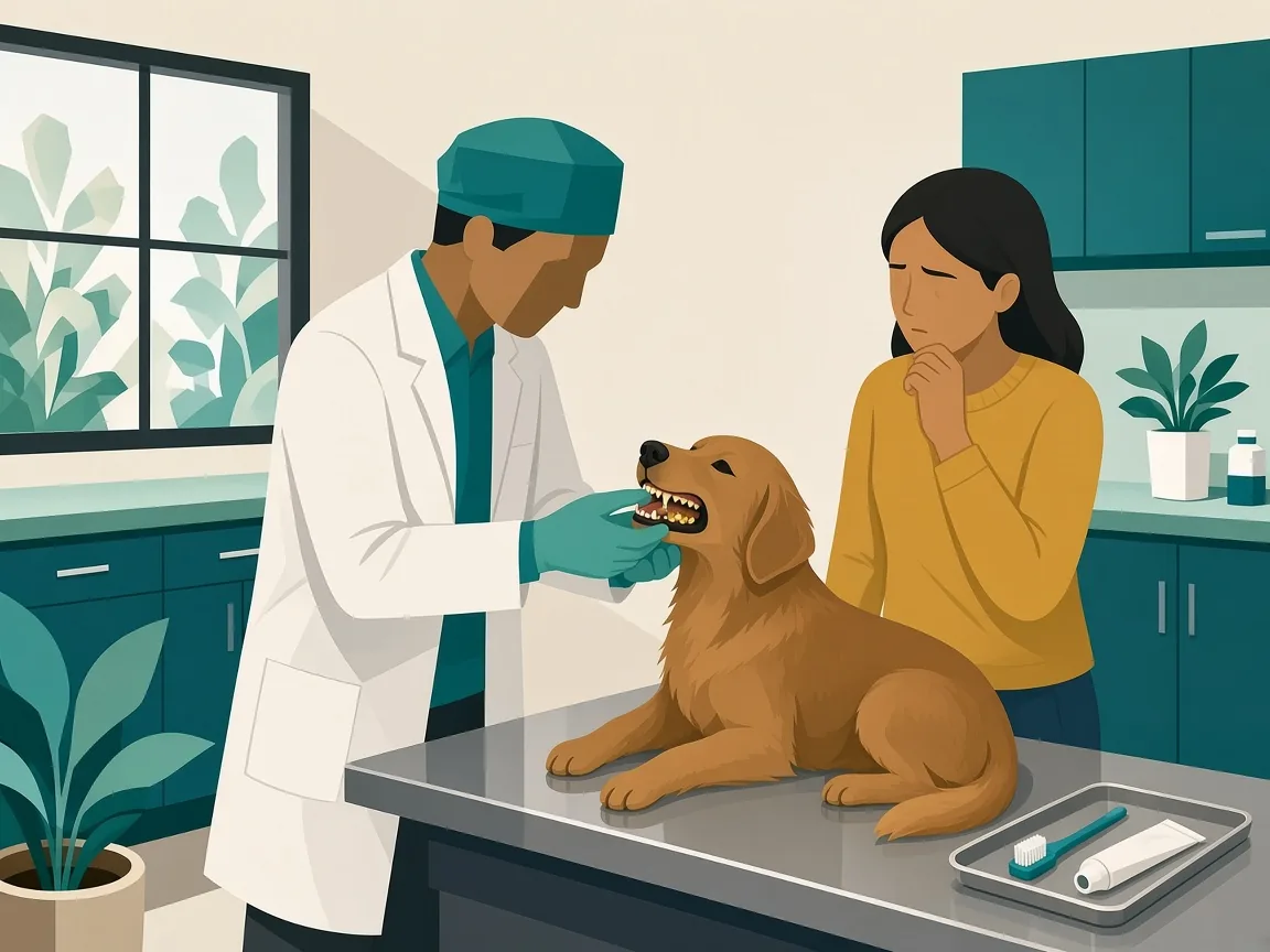
Luna's Teeth Taught Us Everything: Why Dental Health Can't Wait
Warning signs of dental disease in dogs and cats, plus prevention, home care, and professional cleaning tips for Indian pet parents.
Read article →
1 in 10 Dogs Have Heart Disease: Why Early Screening Matters
Heart disease affects 1 in 10 dogs and 1 in 7 cats. Learn why early cardiac screening helps pets live longer, healthier lives.
Read article →
National Pet Day 2026 (April 11): A Day to Reconnect With Your Pet in Ways That Truly Matter
National Pet Day 2026 ideas beyond treats — wellness checks, enrichment, bonding activities, and preventive care for dogs and cats.
Read article →